Team 3 - FOSFO
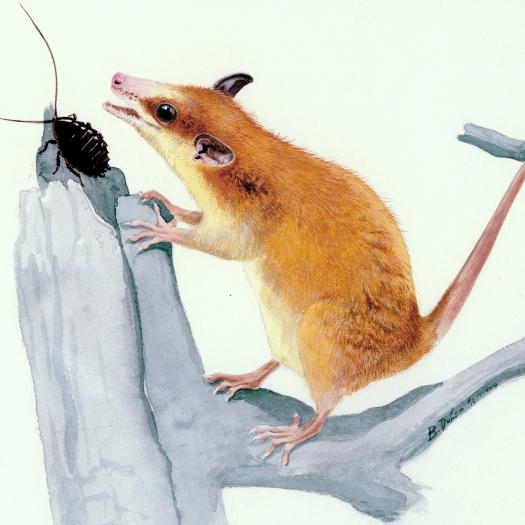

FOSFO - Forms, Structures and Fonctions
The team’s projects cover a broad field of investigation both temporally (almost all of the Phanerozoic) and taxonomically (all vertebrate clades), as well as in terms of studied environments (continental and aquatic).
In the projects carried out by team members emphasis is placed on anatomical, physiological, and ecological aspects of extinct organisms by combining both traditional and innovative methods. The use of extant taxa for comparison is also a strong component of the team’s work.
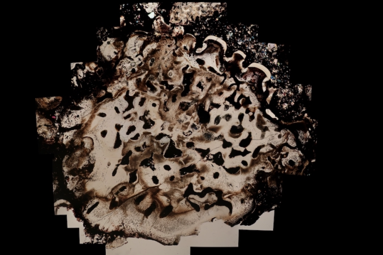
Coupe transversale dans le squelette dermique d'Astraspis (Ordovicien des États Unis) reconnaissable à ses odontodes, composés d'une couche d'émailloide recouvrant de la dentine, qui surplombent une épaisse couche d'os lacunaire. Crédit photo: Damien Germain. (Lemierre & Germain 2019)
ENGLISH Cross-section in the dermal skeleton of Astraspis (Ordovician of the USA) showing odontods, composed of an enameloid layer covering dentine, which overlay a thick cancellous bone layer. Photo credit: Damien Germain. (Lemierre & Germain 2019)

Dessin du crâne de Microcleidus melusinae, un nouveau plésiosaure daté du Toarcien (Jurassique) du Luxembourg. Crédit: Peggy Vincent. (Vincent et al. 2019; https://hal-mnhn.archives-ouvertes.fr/CR2P/hal-02298448v1)
ENGLISH: Draw of the skull of Microcleidus melusinae, a new plesiosaurian from the Toarcian (Jurassic) of Luxembourg. Credit: Peggy Vincent. (Vincent et al. 2019; https://hal-mnhn.archives-ouvertes.fr/CR2P/hal-02298448v1)

Anatomie endocrânienne d'un spécimen de plésiosaure du Crétacé du Maroc. (Allemand et al. 2019; https://hal-mnhn.archives-ouvertes.fr/CR2P/hal-02354078v1)
ENGLISH Endocranial anatomy of a plesiosaur specimen from the Cretaceous of Morocco. (Allemand et al. 2019; https://hal-mnhn.archives-ouvertes.fr/CR2P/hal-02354078v1)
One of the goals of paleontology is to understand the mechanisms involved in the evolution of species. In order to achieve this, palaeontologists must look at these species’ adaptations, including the nature of their ontogenetic development, their anatomical and functional characteristics, and, more generally, their place in paleoecosystems. Thanks to the development of high-performance analytical techniques, this approach has come to play a major role in the functional and ecological study of fossil organisms.
In this context, our team addresses paleobiological issues through three main unifying “temporal projects”, around which revolve other related (sub)projects. The main projects are: Paleozoic terrestrialization (i.e. the colonization of land by animals), terrestrial animals’ secondary return to aquatic life (as soon as the end of the Paleozoic and more extensively during the Mesozoic), and specialized adaptations during the Cenozoic. These major phenomena are studied in order to better understand evolutionary mechanisms and the anatomical and physiological innovations responsible for the emergence and radiation of the vertebrate groups involved. The three areas intersect by the use of various techniques, the most widely used being bone microanatomy and histology, as well as isotope geochemistry, X-ray microtomography, and analytical morphometry. In addition to these three major projects, the team also includes an independent cross-team “historical project”, devoted to the History of Science and involving several members of the team for numerous years.
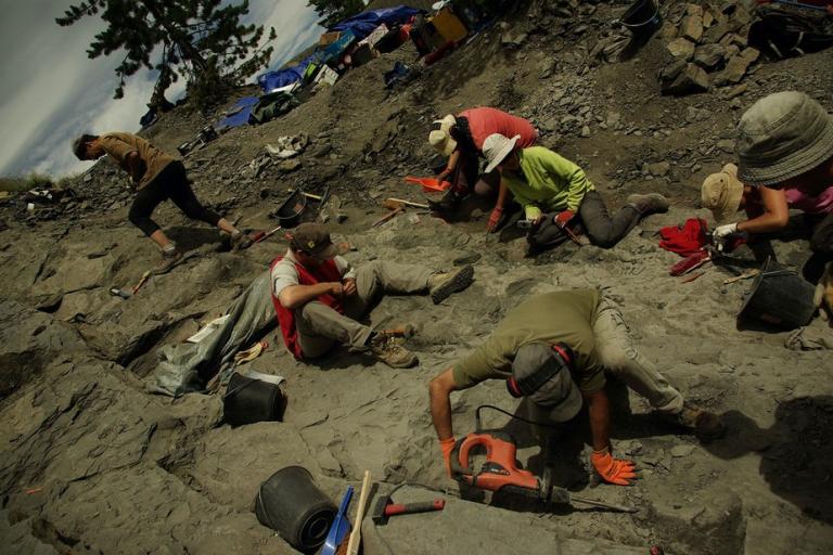
Réserve naturelle géologique de Haute-Provence. Découverte de divers restes de vertébrés du Crétacé dont un squelette partiel de plésiosaure, l'un des plus complet datant de l'Albian en Europe.
Alpes de Haute-Provence. Finding of various vertebrate remains including a partial skeleton of a Cretaceous plesiosaur, one of the most complete Albian plesiosaurian specimens known from Europe.
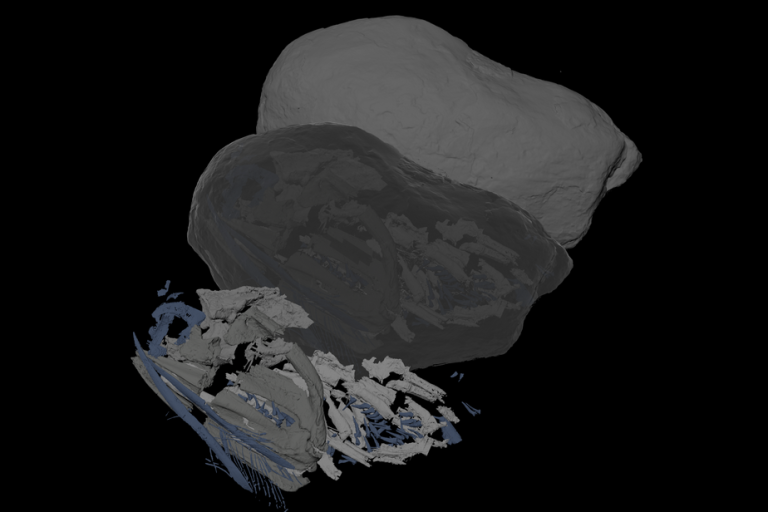
Acanthodes, un poisson cartilagineux de 300 millions d'années, apparenté aux requins et aux raies, visualisé par tomographie aux rayons X.
English: Acanthodes, a 300 million year old cartilaginous fish, a relative of sharks and rays, visualised using computed tomography.
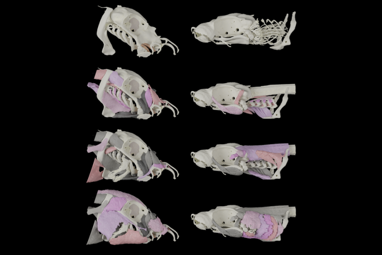
Les muscles de la tête et le squelette de la chimère éléphant (gauche, Callorhinchus milii) et de la petite roussette ( droite, Scyliorhinus canicula), visualisés par tomographie synchrotron aux rayons X.
English: The head muscles and skeleton of a ploughnosed chimaera (Left, Callorhinchus milii) and small-spotted catshark (Right, Scyliorhinus canicula), visualised using x-ray synchrotron tomography.
Researchers and teacher-researchers: Christine Argot (MC MNHN), Nathalie Bardet (DR CNRS), Gaël Clément (PR MNHN), Damien Germain (MC MNHN), Helder Gomes Rodrigues (CR CNRS), Sandrine Ladevèze (CR CNRS), Alain Pradel (MC MNHN), Alexandra Quilhac (MC SU), Jean-Sébastien Steyer (CR CNRS), Peggy Vincent (CR CNRS).
Emeritus teacher-researchers: Marc Godinot, Philippe Janvier, Louise Zylberberg.
Post-doctoral fellows and PhD ongoing: Vincent Decuypere, Guillaume Houée, Antoine Logghe, Nicolas Seon.